
A few decades ago, if doctors wanted a closer look inside the body, they often had to perform surgery. Today, that’s rarely necessary. Thanks to modern medical imaging, doctors can examine organs, bones, blood vessels, and even the brain without making a single incision.
Among the most commonly used imaging techniques are CT (Computed Tomography) and MRI (Magnetic Resonance Imaging) scans. Both allow doctors to see inside the body, but they work in completely different ways and serve different purposes.
How does a CT scan work?
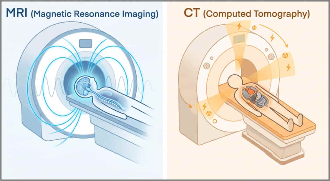
A CT scan uses X-rays to create detailed images of the body’s internal structures. Unlike a standard X-ray, which captures just one image, a CT scanner rotates around the patient, taking hundreds of X-ray pictures from different angles.
A computer then combines these images into thin cross-sectional “slices” of the body, almost like flipping through the pages of a book. These slices can even be assembled into a three-dimensional image, giving doctors a much clearer view of what’s happening inside.
Because CT scans are extremely fast, they are often used in emergency situations. They can quickly detect broken bones, internal bleeding, lung diseases, strokes, and many types of cancer, helping doctors make rapid treatment decisions.
How does an MRI scan work?
An MRI scan doesn’t use X-rays or any other form of ionising radiation. Instead, it relies on a powerful magnetic field and radio waves.
The human body is made up largely of water, and every water molecule contains tiny hydrogen atoms. Inside the MRI scanner, the strong magnetic field temporarily aligns these hydrogen atoms. Short bursts of radio waves then disturb this alignment. As the atoms return to their normal position, they emit tiny signals that are detected by the scanner.
A computer processes these signals to produce highly detailed images, especially of soft tissues that are difficult to see using X-rays.
This makes MRI particularly useful for examining the brain, spinal cord, muscles, ligaments, tendons, cartilage, and other soft tissues. It is also widely used to diagnose neurological disorders, sports injuries, and many joint problems.
Which scan is better?
The answer depends entirely on the medical question.
If doctors need a rapid assessment, for example, after a road accident or when internal bleeding is suspected, a CT scan is usually the preferred choice because it is quick and excellent at imaging bones and blood.
If they need a closer look at soft tissues, nerves, or the brain, an MRI often provides far more detail.
Rather than competing technologies, CT and MRI scans complement each other. Each has unique strengths, and doctors choose the one that provides the most useful information for a particular condition.
Modern imaging has transformed medicine, allowing physicians to diagnose diseases more quickly, accurately, and safely than ever before, all without the need for exploratory surgery.


