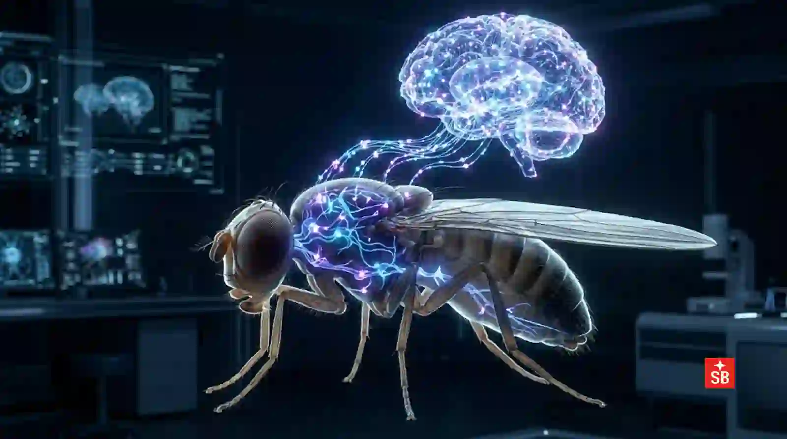
Somewhere in a freezer at Janelia Research Campus, Howard Hughes Medical Institute (HHMI) USA, a single male fruit fly smaller than a grain of rice, with a brain you could balance on the head of a pin, was sliced into thousands of wafer-thin sections, photographed neuron by neuron, and reassembled inside a computer. What came out the other side is something science has never had before: a complete, synapse-by-synapse map of every neuron in an animal’s brain and nervous system, all 166,000 of them, wired together by 125 million connections.
It sounds like a technical footnote. It isn’t. It might be one of the most important maps ever drawn not of a country or a coastline, but of thought itself.
Why a Fly’s Brain Is Everyone’s Business
It’s easy to shrug this off as an insect story. But here’s the thing that should stop you: roughly 75% of the genes known to cause disease in humans have a working counterpart in the fruit fly. When a fly’s neurons misfire, forget, panic, court a mate, or freeze in fear, they’re running on genetic and molecular hardware startlingly similar to ours. We can’t yet map a human brain because 86 billion neurons is still beyond reach, but we can map a fly’s. And a fly, it turns out, is complicated enough to teach us the rules.
Think about what your brain does in a single second without asking your permission: it recognizes a friend’s face in a crowd, remembers where you parked, keeps you upright, and decides whether that noise behind you is dangerous. Every one of those judgments is a signal flowing through a wiring diagram nobody has ever fully seen until now, in a much smaller creature.
What It Actually Took to Build This
This wasn’t a weekend project. It was roughly a decade of work by teams at HHMI Janelia Research Campus, Google Research, Princeton, and a global consortium of labs, built on a simple but staggering process: slice a brain into thousands of ultra-thin sections, image every one under an electron microscope, then use AI to trace each individual neuron: a threadlike structure that can twist and branch thousands of times through the entire stack without ever confusing it with its neighbor. Then, critically, humans checked the machine’s work. Every neuron in this map was proofread by a person.
That human proofreading detail matters more than it might seem. In 2021, only 15% of these neurons had been verified by hand. Getting to 100% took years of painstaking, unglamorous labor: a reminder that even in the age of AI, some of science’s biggest breakthroughs still depend on people willing to double-check a machine, one cell at a time.
The result covers not just the brain but the ventral nerve cord – the fly’s version of a spinal cord. So for the first time, researchers can trace a signal all the way from an eye or antenna, through the brain, down to the muscles that make the fly move. That’s the difference between a map of a city’s streets and a map that also shows you exactly which car takes which road to get home.
The Twist: Male and Female Brains, Compared
What makes this particular map extraordinary is that it’s a male connectome, built to sit alongside an earlier complete map of a female fly brain. For the first time, scientists could compare two brains from the same species, neuron by neuron, and ask: where do male and female brains actually differ?
The answer is more precise than anyone expected. Out of thousands of matched cell types, only a small fraction (around 5% of neurons in males) showed clear sex-specific differences. Most of the brain’s architecture is shared. The differences that do exist cluster in circuits tied to courtship and aggression, controlled in part by two genes, fruitless and doublesex, that had been suspected for years to shape these behaviors but had never been traced through a complete circuit before.
There’s something quietly moving about that finding. It suggests that the vast majority of what builds a brain: the scaffolding of memory, perception, and movement isn’t about sex at all. It’s shared circuitry, present in both, doing the same essential job.
Why This Should Make You Feel Something
We tend to think of neuroscience discoveries as distant and clinical, but this one has a very human undertone. For over a century, the humble fruit fly has been a stand-in for us in the lab: patient, uncomplaining, endlessly reproducing across generations of experiments, quietly helping researchers understand genetics, aging, and now the very architecture of thought.
This connectome is a proof of concept for a promise that once sounded like science fiction: that we might, one day, be able to look at a damaged or diseased human brain circuit and know, precisely, what’s broken and why. The NIH has already called this project a forerunner to larger mammalian brain-mapping efforts. The same imaging and AI pipeline used here is now being pointed at the brains of small fish, testing whether these methods can scale toward a vertebrate brain – a nervous system built more like ours.
We are, in other words, watching the opening chapter of a much longer story: the one where neuroscience stops describing the brain in general terms and starts reading it like a circuit diagram, wire by wire. A fly brain today. Perhaps, within our lifetimes, pieces of a mammalian one. That’s not a small thing to come from an insect most people spend their lives swatting away.
References
Berg, S., Beckett, I. R., Costa, M., Schlegel, P., Januszewski, M., Marin, E. C., Nern, A., Preibisch, S., Qiu, W., Takemura, S., … Rubin, G. M. (2025). Sexual dimorphism in the complete connectome of the Drosophila male central nervous system. bioRxiv. https://doi.org/10.1101/2025.10.09.680999
Dorkenwald, S., Matsliah, A., Sterling, A. R., Schlegel, P., Yu, S.-C., McKellar, C. E., Lin, A., Costa, M., Eichler, K., Yin, Y., … Murthy, M. (2024). Neuronal wiring diagram of an adult brain. Nature, 634, 124–138. https://doi.org/10.1038/s41586-024-07558-y