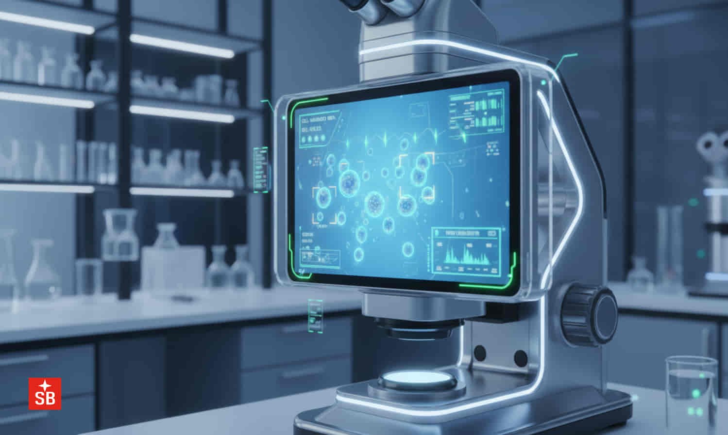
A microscope has long been the doctor’s window into the hidden world of disease. Now, artificial intelligence is turning that window into a wise guide that can quickly spot tiny clues the human eye might miss. It is like giving a microscope a second pair of intelligent eyes that never grow tired. “Show me the smallest hint, and I will search every corner,” the AI seems to say. As we all know, “Many hands make work simple.” In the same way, when doctors and AI work together, diagnosing diseases becomes faster, more consistent, and more reliable. The goal is not to replace doctors, but to help them make better decisions with greater confidence.
Modern healthcare demands speed, accuracy, and fairness. Every minute matters when a patient is waiting for a diagnosis. Traditional microscopy often requires specialists to spend long hours examining hundreds of samples.
AI can rapidly scan digital images, identify suspicious areas, and prioritize cases that need urgent attention. This reduces repetitive work while improving consistency. It is like having a highly alert assistant who whispers, “This sample needs your attention first.” Instead of searching for a needle in a haystack, doctors are guided directly to the most important findings.
One of the greatest strengths of AI integrated digital microscopy is its ability to capture, store, and share high quality images securely. Digital slides can be archived for many years, retrieved instantly, and shared with experts anywhere in the world. This technology supports telepathology, allowing specialists in different cities or countries to examine the same sample without travelling. The microscope no longer works alone. It becomes a bridge connecting laboratories across continents, proving that distance is no longer a barrier to expert medical care.
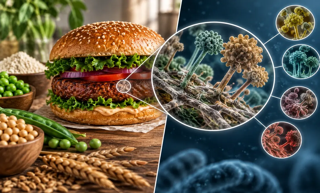
The technology is finding applications across many medical fields. In microbiology, AI helps detect tiny organisms such as malaria parasites, bacteria, fungi, amoebae, and algae from stained samples. In tuberculosis diagnosis, it rapidly identifies acid fast bacilli in sputum smears, reducing the workload of laboratory staff and speeding up reports.
In hematology, it assists in reviewing blood smears, counting different blood cells, and detecting abnormal cells. In cytogenetics, AI simplifies chromosome analysis and karyotyping, making it easier to identify genetic abnormalities. These intelligent systems work like tireless detectives, carefully examining every microscopic clue before raising an alert.
Cancer diagnosis is another area where AI is creating a major impact. During cancer prescreening, AI can identify suspicious cells and tissue changes that deserve closer examination by a pathologist. It also improves immunohistochemistry by measuring biomarker expression more accurately and consistently. Deep learning algorithms and convolutional neural networks, commonly called CNNs, act like experienced pattern hunters.
They learn from thousands of medical images and recognize complex disease patterns with remarkable speed. By combining pathology, radiology, and genomic information, AI supports more personalized treatment decisions. In simple words, it helps doctors choose the right treatment for the right patient at the right time.
Digital microscopy is also becoming valuable in forensic science. High resolution digital images provide reliable documentation that can support criminal investigations and legal evidence. Every microscopic detail is carefully preserved, making records easier to review and share whenever needed. The old saying, “A picture is worth a thousand words,” becomes especially true when a single microscopic image can help solve a medical mystery or support justice in a courtroom.
Despite its enormous promise, AI is not a magic wand. Every intelligent system depends on high quality digital scanners, large collections of accurately labelled images, clinical validation, regulatory approval, and strong cybersecurity. Poor quality samples or unusual cases may confuse even the smartest algorithms. Ironically, a machine built to reduce errors can still make mistakes if it learns from incomplete or imperfect data. That is why expert pathologists continue to play the leading role. AI is an assistant, not the final judge.
The future of disease diagnosis lies in teamwork between human intelligence and artificial intelligence. Doctors bring experience, clinical judgement, and compassion, while AI contributes speed, precision, and consistency. Together they form a powerful partnership that can improve patient care, laboratory efficiency, and access to expert diagnosis across the world. The microscope is no longer just an instrument. It is becoming a smart storyteller, quietly revealing the hidden secrets of disease, one cell at a time.