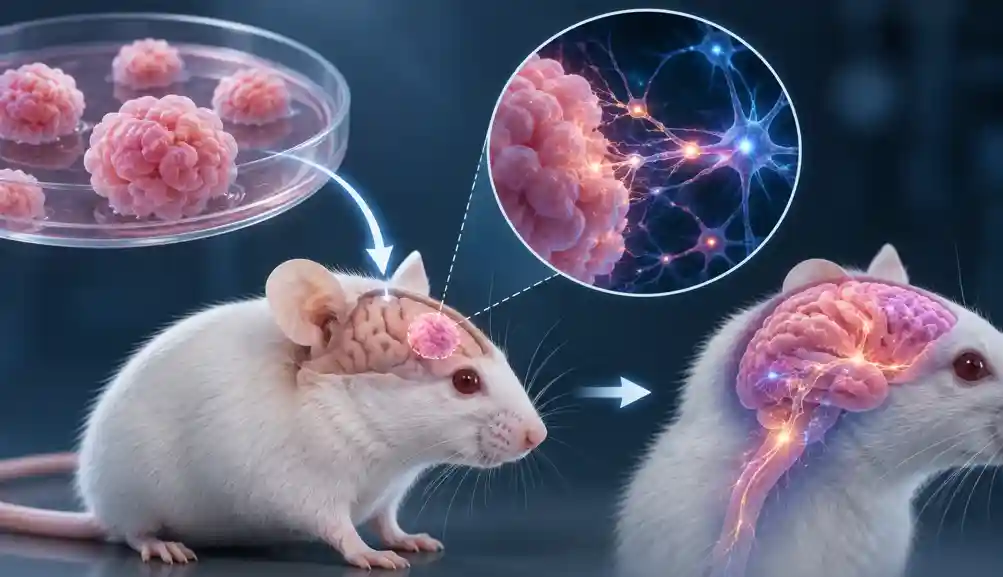
Scientists have now taken a remarkable step toward answering that question. Researchers at Stanford University transplanted human brain organoids miniature, lab-grown pieces of human cortical tissue into genetically engineered mice whose cerebral cortex was largely absent. Instead of simply surviving, the human tissue grew, matured and formed extensive connections with the mouse nervous system.
Why put human brain tissue into mice?
Brain organoids are usually grown in laboratory dishes. They can reproduce certain aspects of human brain development, but they lack many of the signals provided by a living organism, including blood supply, sensory input and connections with other organs.
The researchers therefore created mice with most of their cerebral cortex missing, leaving physical space for transplanted human tissue to develop.
The result was striking.
Over several months, the human tissue expanded nearly fivefold and eventually occupied more than 90% of the available cortical space. The transplanted tissue developed multiple types of human neurons and formed connections extending into the mouse brain and even toward the spinal cord.
Something scientists had struggled to grow appeared
Among the cells that developed were von Economo neurons, specialized neurons associated with complex social and cognitive functions and implicated in certain neurodegenerative diseases.
These cells have been extremely difficult to reproduce in conventional laboratory cultures. Their appearance in the transplanted tissue suggests that the living brain environment provides developmental signals that organoids cannot fully reproduce in a dish.
Did the mice become smarter?
No.
Despite containing a large amount of human cortical tissue, the animals did not suddenly develop human-like intelligence or consciousness. Behavioural testing showed that the mice remained broadly similar to normal animals, although the transplanted human tissue did appear to contribute to some motor and memory functions.
The human tissue also did not develop into a complete human cerebral cortex. It lacked some of the organized layering characteristic of a normal human cortex.
Why does this matter?
The real purpose is medicine.
The researchers used the model to investigate how human brain tissue responds to conditions such as oxygen deprivation, which can cause neurological damage. The human-derived tissue showed vulnerabilities that differed from those seen in ordinary mice, potentially making the model useful for studying disorders such as cerebral palsy, epilepsy, autism and other neurodevelopmental conditions.
The experiment also raises important ethical questions. As human neural tissue becomes increasingly integrated into animals, researchers must consider how much human brain function can safely be introduced into an animal and where appropriate ethical boundaries should lie. The researchers used dedicated ethical oversight and deliberately limited the extent and duration of the experiments.
This is therefore not the creation of a “human-brain mouse.”
It is something scientifically more useful: a living experimental system in which human brain tissue can be studied inside a functioning organism.
And that could give scientists something they have never had before—a much closer look at how the uniquely human brain develops, malfunctions and responds to potential treatments.
Sources:
https://med.stanford.edu/news/all-news/2026/09/model-to-study-brain-development.html