Electronic cigarettes are often presented as a safer alternative to traditional cigarettes, but new research suggests that their vapour may still disturb important processes inside our cells. In simple words, vaping may not be as harmless as it looks. Like a quiet spark that can start a much larger fire, e-cigarette exposure may trigger cellular changes that help damaged cells survive. A team of molecular biologists from the National Institute of Molecular Biology and Biotechnology (NIMBB) at the University of the Philippines Diliman (UPD) College of Science investigated whether electronic cigarettes alter cellular and molecular pathways associated with oncogenesis.
Background & Research Motivation
Electronic cigarettes are frequently marketed as safer alternatives to combustible tobacco because of their reduced chemical load. However, “less harmful” does not necessarily mean “harmless”, and this distinction is important when studying long-term health effects. Earlier work by Dr. Reynaldo Garcia, published in Scientific Reports, identified that combustible cigarette smoke activates a long non-coding RNA (lncRNA) called SCAL1, or smoke- and cancer-associated lncRNA transcript 1. SCAL1 detoxifies reactive oxygen species (ROS), which creates a survival advantage. Rather than undergoing apoptosis, or programmed cell death, damaged cells survive and accumulate oncogenic potential. In this situation, the cell’s protective shield may become a double-edged sword.

The researchers therefore sought to determine whether e-cigarette aerosol triggers a similar cellular survival mechanism. The question was simple but important: if cigarette smoke can activate a cancer-associated survival pathway, can vape exposure do something similar?
Key Findings & Mechanisms
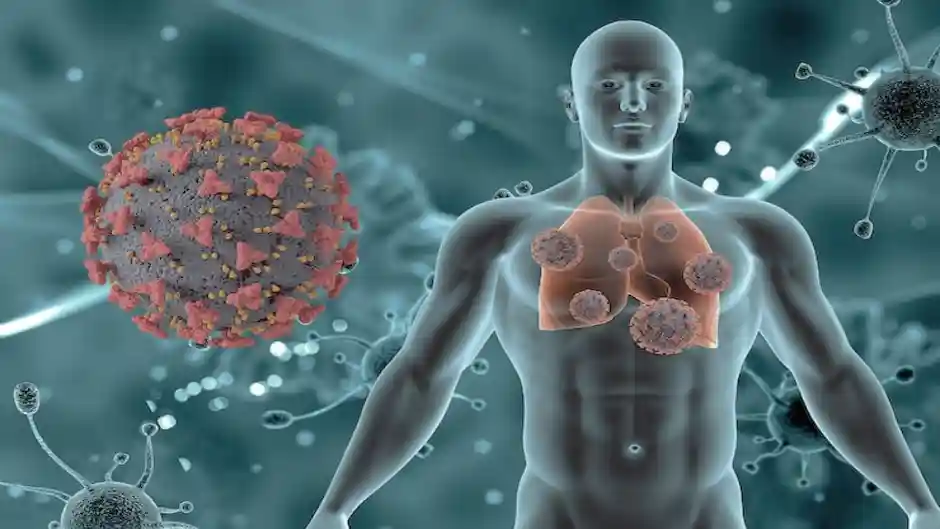
The researchers identified and characterized an e-cigarette-responsive long non-coding RNA, naming it VALT1, or vape-associated lncRNA transcript 1. Public RNA-sequencing data showed that VALT1 was elevated both in the oral epithelia of vape users and in non-small cell lung cancer (NSCLC) tissues. In other words, this molecular signal appeared in both vape-exposed tissue and a major form of lung cancer, raising an important biological question.
Using cell models, including the human lung carcinoma cell line A549 and non-tumorigenic bronchial epithelial cells BEAS-2B, the team observed several distinct cellular responses. These findings provide a molecular picture of how vaping may influence cellular behaviour.
Induction of VALT1
Exposure to e-cigarette smoke extract (eCSE) caused a dose-dependent increase in cytoplasmic VALT1 expression. This means that as the exposure increased, VALT1 levels also increased. The relationship works almost like turning up a molecular volume knob, with stronger exposure producing a stronger response.

Tumorigenic Hallmarks
Elevated VALT1 triggered phenotypes typical of transformed cancer cells, including accelerated cell proliferation, increased cellular migration, enhanced ROS detoxification, cytoskeletal disorganization, and nuclear remodeling. These are important tumorigenic hallmarks because cancer cells do not simply multiply, they change their behaviour, movement, structure, and ability to withstand cellular stress.
Resistance to Programmed Cell Death
VALT1 promoted resistance to cytotoxic stress and prevented apoptosis, allowing genetically stressed lung cancer cells to persist rather than die off. Normally, apoptosis acts like a cellular quality-control system, removing severely damaged cells from the body. When this safeguard is weakened, damaged cells may get a second chance to survive. As the old saying goes, “A small leak can sink a great ship”, and in cellular biology, even a small change in a protective pathway may have wider consequences.
Autonomous Activity
Overexpressing VALT1 artificially produced these pro-tumorigenic hallmarks even without direct exposure to vape liquid, while knocking down VALT1 using siRNA attenuated the malignant features. This is an important observation because it suggests that VALT1 is not merely a passive marker of exposure. It may actively participate in producing the observed cellular changes.
In simple terms, the researchers used a molecular “switch test”. When VALT1 was increased, cancer-like characteristics appeared, and when VALT1 was reduced, those characteristics became weaker. This provides stronger evidence for a functional role of the lncRNA.
Implications and Conclusions
The authors concluded that e-cigarettes are not physiologically harmless. VALT1 acts as an active mediator through which e-cigarette vapor induces transformed, cancer-like behaviours in lung cells even in the absence of traditional driver mutations. This finding adds another layer to the discussion about vaping and cancer biology.
The study also highlights the growing importance of non-coding RNA biology. DNA may provide the basic instructions of life, but non-coding RNAs can influence how those instructions are regulated. In this sense, the genome is not just a book of genes, it is also a complex network of molecular switches and signals.
While lung cancer typically requires decades of latency to manifest clinically, these findings establish that e-cigarette vapor directly dysregulates non-coding RNA transcriptomes and initiates molecular pathways that support tumor survival and progression. However, cellular and molecular findings should not be interpreted as proof that vaping directly causes lung cancer in humans. Further animal studies, long-term epidemiological studies, and clinical research are needed to establish the full health consequences.
The message from this research is therefore clear: vaping may change more than what meets the eye. Beneath the visible vapour, a complex molecular story may be unfolding inside cells. As the saying goes, “Prevention is better than cure”, and understanding these early molecular changes may help researchers better assess the long-term risks of e-cigarette exposure.
Source:
Mirador, D. A. R., Ferrer, J. L. M., Lin, K. D. H., & Garcia, R. L. (2026). Vape-Associated lncRNA Transcript 1 (VALT1) Amplifies the Tumorigenic Effects of e-Cigarette Vapor in Lung Epithelial Cells.