Radioimmunoassay (RIA) is a highly sensitive laboratory technique used to measure extremely small amounts of substances in biological samples. It is especially useful for detecting hormones, drugs, vitamins, and other substances that may be present in very low concentrations. RIA was developed by Rosalyn Yalow and Solomon Berson in 1959 and became an important method in clinical diagnosis and medical research. The technique combines the specificity of an antigen–antibody reaction with the high sensitivity of radioactive detection.
Principle
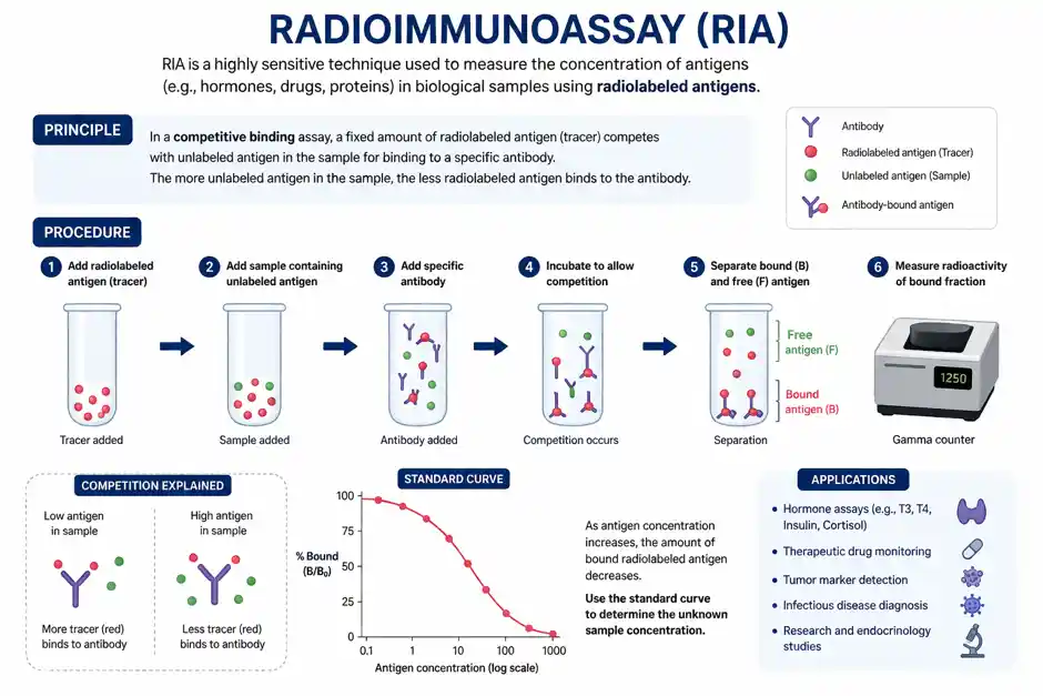
The basic principle of RIA is competitive binding. In this method, a radiolabelled antigen and the unlabelled antigen present in the patient’s sample compete for a limited number of binding sites on a specific antibody.
A known amount of radioactive antigen is mixed with a specific antibody and the sample containing an unknown amount of antigen. Both labelled and unlabelled antigens compete to bind with the available antibodies. If the sample contains a higher amount of unlabelled antigen, it will occupy more antibody-binding sites, leaving less radioactive antigen bound to the antibody.
After the reaction is complete, the antigen bound to the antibody is separated from the free antigen. The radioactivity is then measured using a suitable radiation counter. In most RIA methods, iodine-125 (¹²⁵I) is commonly used as the radioactive label and its activity can be measured using a gamma counter.
The amount of radioactivity measured is inversely related to the concentration of antigen in the sample. A standard curve prepared using known antigen concentrations is used to determine the concentration of the unknown sample.
Procedure
The procedure of RIA can be summarized in the following steps:
- Preparation of reagents: A specific antibody, radiolabelled antigen, unlabelled antigen standards, and suitable buffer are prepared.
- Addition of samples: Known standards and unknown patient samples are placed into separate tubes.
- Addition of antibody: A fixed amount of specific antibody is added to each tube.
- Addition of labelled antigen: A known amount of radiolabelled antigen is added.
- Incubation: The mixtures are incubated under suitable conditions to allow the antigen and antibody to interact.
- Separation: The antigen bound to the antibody is separated from the unbound antigen.
- Measurement: The radioactivity of the appropriate fraction is measured using a radiation counter.
- Interpretation: The results are compared with a standard curve to calculate the concentration of the substance in the unknown sample.
Applications
RIA has been widely used in clinical laboratories and biomedical research because of its high sensitivity. It is particularly useful for measuring hormones such as insulin, thyroid hormones, growth hormone, cortisol, and reproductive hormones. It has also been used to measure vitamins, steroid hormones, therapeutic drugs, and certain antigens.
RIA has helped in the diagnosis and study of conditions involving the thyroid, pancreas, adrenal glands, reproductive system, and growth disorders. Researchers have also used it to study hormone secretion, drug metabolism, and other biological processes.
Although RIA is highly sensitive and specific, it has some disadvantages. It requires radioactive substances, specialized equipment, trained personnel, radiation-safety precautions, and proper disposal of radioactive waste. Because of these limitations, many RIA procedures have been replaced by ELISA, chemiluminescent immunoassays, and other non-radioactive techniques.
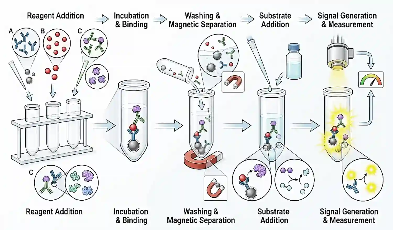
Even so, RIA remains an important milestone in diagnostic medicine. Its development demonstrated how immunological reactions could be combined with sensitive detection methods to measure substances present in extremely small quantities.