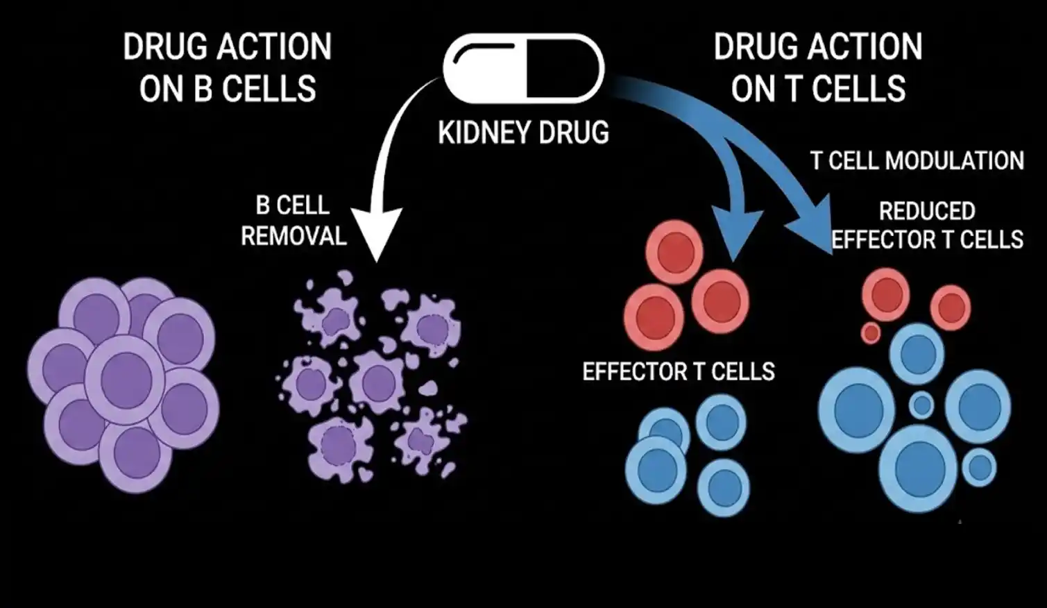
A drug designed to remove one type of immune cell may be changing another and that could help explain why it works for some kidney patients but not others. Researchers at Nagoya University have found that rituximab, a drug that removes B cells from the blood, can trigger major changes in T cells in people with difficult-to-treat nephrotic syndrome. In patients who responded well to treatment, their T cells showed signs of improved energy production and reduced cellular stress.
To understand why this matters, first let’s consider what happens in nephrotic syndrome. The kidneys contain specialised filtering structures that prevent large proteins from escaping into the urine. When these filters become damaged, large amounts of protein can leak out, causing the characteristic protein loss seen in nephrotic syndrome.
One common form is minimal change disease (MCD). Under an ordinary microscope, the kidneys can appear almost normal. But at much higher magnification, the tiny extensions of specialised filtering cells called podocytes become flattened, disrupting the filtration barrier.

Steroids are commonly used to control the disease. However, some adults become dependent on steroids, meaning the disease returns when the dose is reduced. Long-term steroid treatment can also cause serious side effects. This is where rituximab enters the picture. Rituximab is an antibody that targets CD20, a protein found on B cells. By eliminating these cells from the circulation, it can help some patients achieve remission. But there has always been a puzzle.
If rituximab works mainly by removing B cells, why do some patients remain in remission even after their B cells return?
The new study provides a possible answer. They collected blood samples from 14 adults with minimal change disease who had been dependent on steroids for about a decade. Samples were examined before rituximab treatment and one month afterward. Six patients were studied using single-cell gene-expression analysis, while another eight were assessed for oxidative stress. They divided the patients into responders and non-responders.
In the responders, T cells underwent substantial changes after treatment. Genes involved in mitochondrial energy production became more active, suggesting that these cells were improving their ability to generate energy. They also observed changes in reactive oxygen species (ROS). ROS are chemically reactive molecules produced during normal cellular metabolism. Small amounts can have useful signalling roles, but excessive ROS can cause oxidative stress and damage cellular components.
The responding patients showed a reduction in T-cell oxidative stress after rituximab treatment. They also had fewer exhausted T cells, which are immune cells that have become functionally weakened after prolonged stimulation. A subset called CD4⁺ cytotoxic T cells showed particularly notable changes, including expansion of certain clones and stronger oxidative-phosphorylation signatures. The non-responders were different. Although rituximab successfully depleted their B cells too, their T cells showed much less of this downstream change.
That suggests something important: removing B cells may be only the first step. What happens to the remaining immune system afterward could influence whether the treatment actually works. They believe these early changes in T-cell metabolism and oxidative stress could eventually become biomarkers that help predict which patients are likely to respond to rituximab.

This was a small observational study. The single-cell analysis involved only three responders and three non-responders, with additional patients included in the oxidative-stress analysis. Larger studies are needed to determine whether these T-cell changes can reliably predict treatment response. For now, the study changes the way scientists think about rituximab. It may look like a B-cell-targeting drug on paper. But its success could depend partly on what happens to the T cells left behind.
Sources: