Part A: TRIzol RNA Isolation
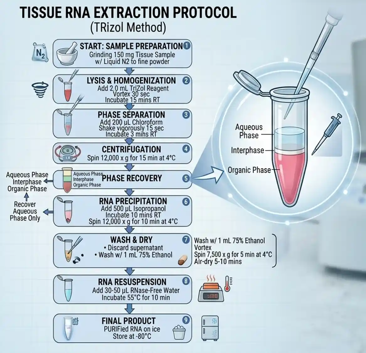
- Homogenisation: Grind 150 mg of tissue into a fine powder using liquid nitrogen.
- Lysis: Add 2 mL of TRIzol reagent to the powdered tissue.
- Incubation: Incubate at room temperature for 5 to 10 minutes to dissociate nucleoprotein complexes.
- Aliquot: Split the lysate equally into two sterile 1.5 mL microcentrifuge tubes (1 mL each).
- Phase Separation: Add 200 µL of chloroform to each tube.
- Agitation: Shake tubes vigorously by hand for 15 seconds.
- Settling: Incubate at room temperature for 5 to 10 minutes.
- Centrifugation I: Spin at 12,000 rpm for 8 minutes at 4 °C.
- Precipitation: Transfer the clear upper aqueous phase to a clean tube.
- Alcohol Addition: Add 500 µL of isopropanol to the collected aqueous phase.
- Chilling: Incubate at -20 °C for 30 minutes to precipitate RNA.
- Centrifugation II: Spin at 10,000 rpm for 8 minutes at 4 °C.
- Washing: Discard supernatant and wash the pellet with 1 mL of 70% ethanol.
- Drying: Centrifuge briefly, remove ethanol, and air-dry the pellet for 5–10 minutes.
- Dissolution: Dissolve the RNA pellet in 20–30 µL of RNase-free water.
Part B: Formaldehyde Gel Electrophoresis
- Gel Casting: Melt 1.2 g of Agarose in 90 mL of RNase-free water.
- Buffer Addition: Cool to 60 °C, then add 10 mL of 10X FA Gel Buffer.
- Pouring: Pour into a casting tray and allow it to solidify.
- Equilibration: Submerge the gel in 1X FA Gel Running Buffer for at least 30 minutes.
- Sample Prep: Mix RNA samples with 5X RNA Loading Buffer at a 4:1 ratio.
- Denaturation: Heat samples at 65 °C for 3–5 minutes, then chill immediately on ice.
- Running: Load samples and run the gel at 5–7 V/cm until tracking dye migrates 2/3 down.
Required Chemicals & Reagents
Core Isolation Reagents
- TRIzol Reagent: Lysis agent containing phenol and guanidinium thiocyanate.
- Chloroform: Induces phase separation (pure chloroform, no isoamyl alcohol added).
- Isopropanol: Precipitates RNA from the aqueous layer.
- Ethanol (70%): Prepared with DEPC-treated water for pellet washing.
- Liquid Nitrogen: Cryogenic grinding of tough tissue samples.
- RNase-free Water: Ultra-pure water treated with 0.1% DEPC and autoclaved.
Electrophoresis Reagents
- Agarose: Standard molecular biology grade for gel matrix matrix construction.
- MOPS (3-(N-morpholino)propanesulfonic acid): Buffering agent for the 10X FA buffer.
- Sodium Acetate: Ionic component for electrophoresis.
- EDTA (pH 8.0): Chelates divalent cations to inhibit remaining RNases.
- Formaldehyde (37%): Denatures RNA secondary structures during the run.
- Formamide: Powerful denaturant included in the loading buffer.
- Glycerol (100%): Provides sample density for well loading.
- Bromophenol Blue: Tracking dye for visual monitoring of migration.
Buffer and Gel Compounding
1. 10x FA Gel Buffer
- 200 mM MOPS
- 50 mM Sodium Acetate
- 10 mM EDTA
2. 1x FA Gel Running Buffer (1 Litre)
- 10X FA Gel Buffer: 100 mL
- 37% Formaldehyde: 20 mL
- RNase-free Water: 880 mL
3. 5x RNA Loading Buffer (12 mL Total Volume)
- 10X FA Gel Buffer: 4 µL
- 37% Formaldehyde: 720 µL
- Formamide: 1184 µL
- 500 mM EDTA (pH 8.0): 80 µL
- Bromophenol Blue solution: 16 µL
- 100% Glycerol: 2 µL
- RNase-free Water: 10 mL
- Storage: Stable for approximately 3 months at 4 °C.
Required Precautions
RNase Decontamination
- Surface Cleansing: Wipe all pipettes, benches, and equipment with RNase-away or 70% ethanol.
- Consumables: Use only certified RNase-free, sterile plasticware and barrier filter pipette tips.
- Water Treatment: Treat all non-certified water with 0.1% DEPC for 12 hours, then autoclave.
Chemical Safety
- Fume Hood Use: Perform all steps involving TRIzol, Chloroform, and Formaldehyde inside a fume hood.
- Personal Protective Equipment: Wear a laboratory coat, safety goggles, and double-gloved Nitrile gloves.
- Phenol Burns: Keep PEG (Polyethylene glycol) nearby to treat accidental TRIzol skin contact.
Troubleshooting Guide
| Symptom / Observation | Potential Root Cause | Corrective Action / Resolution |
| Low RNA Yield | Incomplete tissue homogenisation. | Grind tissue into a completely fine powder; do not allow it to thaw during crushing. |
| Low RNA Yield | Incomplete dissolution of the pellet. | Incubate RNA at 55–60 °C for 10 minutes in RNase-free water to completely dissolve. |
| A260/A280 Ratio < 1.6 | Phenol or protein contamination. | Do not touch the interphase layer when removing the upper aqueous phase. |
| A260/A230 Ratio < 2.0 | Organic solvent carryover. | Ensure the 70% ethanol wash step is thorough; completely air-dry the pellet. |
| Degraded RNA / Smeared Gel | Endogenous or exogenous RNase activity. | Work faster during tissue lysis; ensure all reagents and equipment are RNase-free. |
| Genomic DNA Contamination | Too much starting tissue used. | Reduce the starting tissue weight or scale up the volume of TRIzol used. |
References
- Chomczynski, P., & Sacchi, N. (1987). Single-step method of RNA isolation by acid guanidinium thiocyanate-phenol-chloroform extraction. Analytical Biochemistry, 162(1), 156-159.
- Rio, D. C., Ares, M., Hannon, G. J., & Nilsen, T. W. (2010). Purification of RNA using TRIzol reagent. Cold Spring Harbor Protocols, 2010(6), pdb-prot5439.
- Sambrook, J., & Russell, D. W. (2001). Molecular Cloning: A Laboratory Manual (3rd ed.). Cold Spring Harbor Laboratory Press.