Phase 1: Pre-Assembly Setup and Tool Decontamination
1. Personal Protective Equipment (PPE)
Put on some clean nitrile gloves, a lab coat, and chemical splash goggles.
2. Reagents Required
The Whatman GF/B or GF/F glass microfibre filters are made from 100% borosilicate glass sheets.
0.1 M Sodium Hydroxide (NaOH): Make fresh for matrix sterilization.
Water free of nucleases: this water has been certified as deionized and free of both RNases and DNases.
Ethanol of 100% molecular biology grade: high-purity absolute ethanol.
An example of a commercial RNase decontamination spray would be RNase Away or RNaseZap.
3. Equipment and Hardware
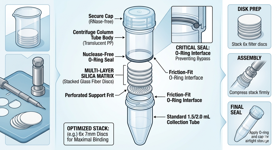
Standard microcentrifuge spin-column housings together with matching plastic internal retaining rings (O-rings).
The metal biopsy punch has a diameter of either 7 mm or 8 mm and is precisely calibrated to match the inner diameter of your column housings.
The fine-tip stainless steel tweezers are intended for use in handling the glass discs.
The benchtop microcentrifuge has a rating of at least 16,000 times g.
As a clean cutting mat, the sterile Petri dish serves this purpose.
4. Environment Sterilization
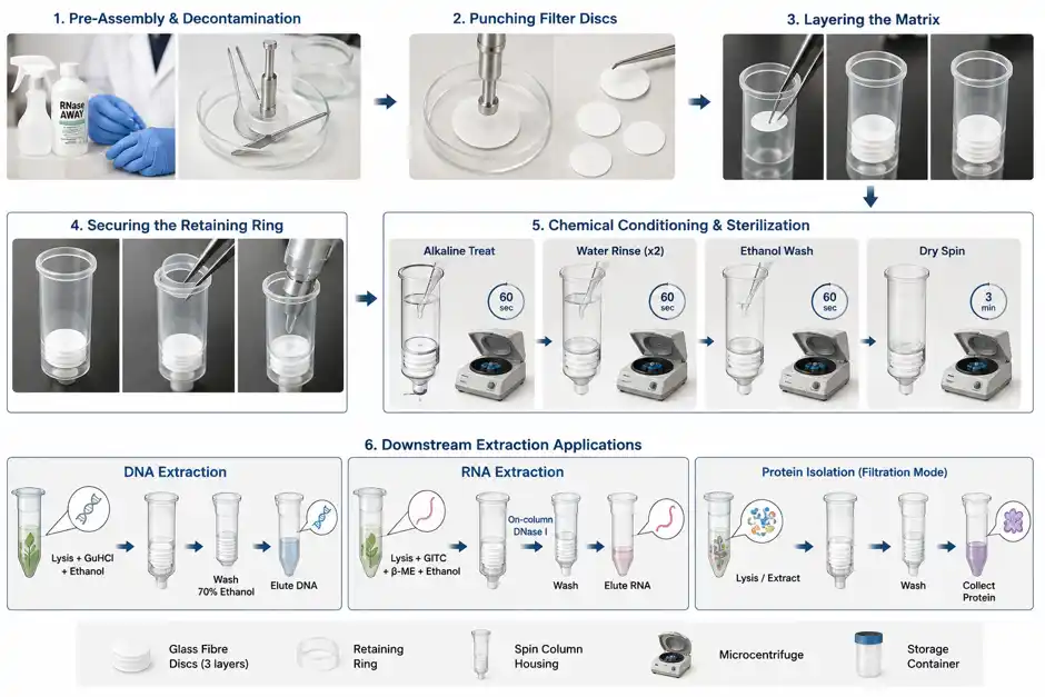
Spray the whole surface of your bench and the pipettes thoroughly with the RNase decontamination solution and then wipe them dry using lint-free wipes.
Put the metal biopsy punch, the fine tweezers, and the plastic cutting dish into 0.1 M NaOH and leave them there for 5 minutes, then rinse them thoroughly with nuclease-free water and place them on a sterile workspace drape.
Phase 2: Mechanical Matrix Assembly
5. Punching Filter Discs
Place a new sheet of Whatman glass fiber filter paper flat inside the decontaminated Petri dish.
Make sure that the biopsy punch is held vertically against the filter sheet, then press down firmly and twist gently in order to cut a circular disc; carry out this procedure again to obtain the required number of discs.
6. Layering the Matrix
Put an empty column housing upright into a sterile microcentrifuge tube rack.
Take the sterile tweezers and pick up one glass fibre disc, then put it flat into the bottom of the column housing.
To the first disc add two more discs directly above it in order to form a uniform 3-layer glass fiber matrix. Note: three layers prevent membrane blowout when the centrifugation is carried out at high speed and optimize the chemical binding capacity.
7. Securing the Retaining Ring
Place the plastic internal retaining ring into the column with the help of your tweezers.
Put the flat rear portion of a sterile 1000 µL pipette tip against the ring, then press it down vertically and firmly so that the ring lies flat against the filter layers. Make sure that there are no warped edges or gaps along the column wall.
Phase 3: Chemical Conditioning and Sterilization
8. Alkaline Treat
Put the assembled column into a clean 2 mL collection tube and pipette 500 µL of 0.1 M NaOH directly onto the centre of the glass matrix.
Place the column assembly in the centrifuge and rotate it at 10,000 times the force of gravity for 60 seconds. Then take it out of the centrifuge and get rid of the caustic flow-through.
9. Hydration Rinses
Put 500 µL of nuclease-free water on to the membrane and then centrifuge it at 10,000 × g for 60 seconds before throwing away the flow-through.
To carry out the water wash step once more, make certain that all the residual NaOH has been fully removed from the matrix.
10. Preparing the Sample for Desiccation and Storage
Put 500 µL of 100% molecular biology grade ethanol on to the filters. Spin them at 12,000 × g for 60 seconds in order to dehydrate and disinfect the silica strands. Throw away the flow-through.
Put the empty column back into the collection tube and spin it at maximum speed (14,000–16,000 × g) for 3 minutes in order to dry the matrix and eliminate all traces of volatile ethanol.
Make sure that the filter membrane is even, opaque and white, and is securely fastened. Put the finished columns into a sterile, airtight container, label the container with the date on which the procedure was carried out and the type of filter, and store them at room temperature.
Phase 4: Downstream Extraction Chemistry Protocols
The fabricated column treats DNA, RNA, and protein through completely different biochemical reactions:
11. Procedure for DNA Extraction (using a chaotropic bridge)
Lysis/Binding Step: To lyse your cell sample, use a buffer containing Guanidine Hydrochloride (GuHCl). Add absolute ethanol so that the final concentration is between 30 and 50 per cent and then load it onto your homemade column. Due to the high concentration of salt the DNA phosphate backbone is dehydrated and is thus made to bind selectively to the silica fibers.
For the washing step, wash the sample using a buffer that contains 70% ethanol in order to remove the cellular salts, proteins, and metabolites, while at the same time leaving the DNA attached.
For the elution step, elute the genomic or plasmid DNA cleanly by using a low-salt buffer (for example, 10 mM Tris-HCl at pH 8.5) or warm nuclease-free water.
12. Procedure for RNA Extraction (Giving the molecule a hydrophobic character)
For the lysis and binding step, lyse the tissue samples in a buffer containing a high proportion of Guanidine Thiocyanate (GITC) and β-mercaptoethanol so as to immediately denature the active RNase enzymes. Then, mix the lysate with an equal volume of 70% ethanol and pass it through the homemade column.
Due to the fact that silica matrices are unable to distinguish between DNA and RNA by themselves, it is necessary to apply a DNase I enzyme mixture directly to the membrane and allow it to act for 15 minutes in order to degrade the genomic DNA that has been co-purified.
For the washing and elution step, wash using a high-ethanol buffer, dry by means of a high-speed centrifugation step, and then elute the pure total RNA by using cold nuclease-free water.
13. Protein Isolation Protocol (Filtration Mode)
1. Prepare all needed solutions and equipment before starting.
2. Place the sample in a clean container. Add the extraction buffer. Mix well to break up the tissue and release proteins.
3. Centrifuge the sample at 10,000 x g for 10 minutes. Collect the supernatant. Throw away the pellet.
4. Set up the filtration unit with the right membrane for your protein size.
5. Pour the supernatant into the filtration unit. Start the filtration using a vacuum or gentle pressure.
6. Wash the membrane with the wash buffer to remove unwanted substances.
7. Collect the filtered protein solution. Store it on ice or at -20°C until you need it.
8. Clean all equipment after use. Dispose of waste following lab safety rules.
Another possible use of the columns is that borosilicate glass matrices are not efficient in using chaotropic bridges to bind soluble native proteins; they should instead be used as clarification or filtration devices.
Procedure: To begin, mix your protein sample with precipitation reagents such as trichloroacetic acid (TCA) or cold acetone. Place the resulting cloudy mixture on the column. The three-layer glass fiber matrix will physically retain the precipitated protein aggregates while permitting the salts and solvent waste to pass smoothly into the collection tube. The pellet that has been trapped can then be washed, air-dried, and resuspended directly on the column by using an SDS-PAGE loading buffer.