Stockholm, October 5, 2026
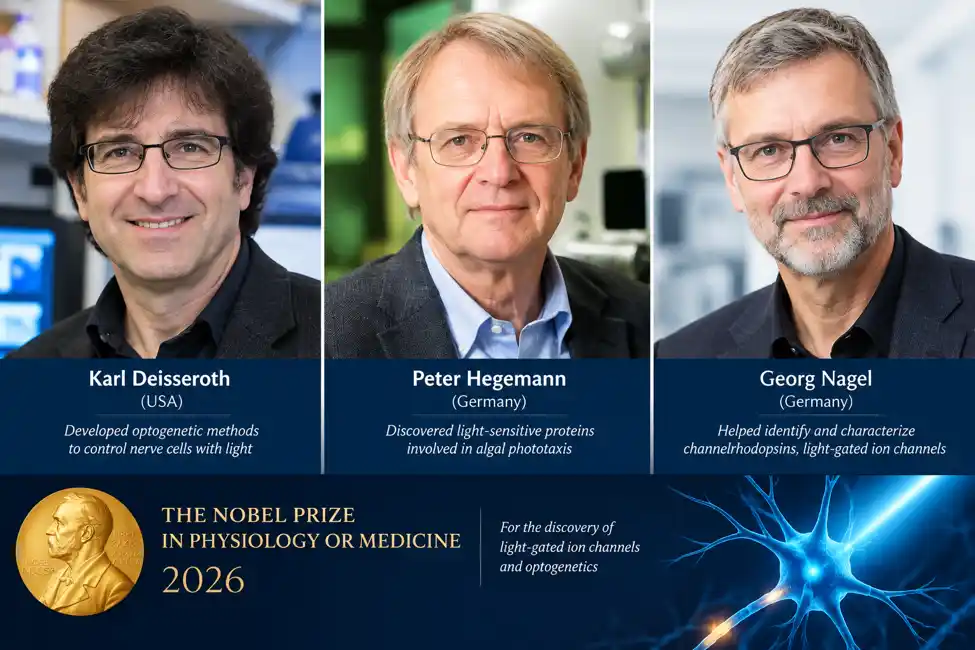
Scientists won the 2026 Nobel Prize in Physiology or Medicine for developing optogenetics, a gene-therapy based technique that uses light to control individual nerve cells in the brain. The Nobel Assembly cited their “discoveries concerning light-gated ion channels and optogenetics.” The three share the 12 million Swedish Kronor prize equally.
The problem: the brain is hard to study
The human brain contains roughly 86 billion neurons. These cells talk to each other with tiny electrical signals, and different types do different jobs. Some help you feel hunger, some form memories, and some control movement. For decades, scientists could only crudely influence them. Electrodes stimulate every cell near their tip, and drugs spread through the whole brain. Neither can single out one type of cell at a specific moment.
In 1979, the Nobel laureate Francis Crick pointed out this limitation and suggested that light might be the answer, because light can be switched on and off in milliseconds and aimed precisely. The Nobel committee noted that this year’s work builds on that idea. Nobody knew how to make a neuron respond to light.
Step one: a clue from pond algae
The answer came from a tiny green alga called Chlamydomonas, which swims toward light, the way a plant leans toward a window. Researchers had long suspected that a light-sensing, rhodopsin-like molecule was involved. In the early 1990s, Hegemann studied how this alga reacts to light within about half a millisecond, and he suspected that a single protein both captures light and acts as an ion channel.
An ion channel is a microscopic gate in a cell’s membrane. When it opens, charged particles (ions) rush in or out, and this is how neurons generate electrical signals. Hegemann’s hunch was that the alga had a gate that opens directly when light hits it.
Step two: finding the gene

Hegemann and Nagel worked together, with Nagel’s expertise in membrane electrophysiology and the Max Planck Institute of Biophysics in Frankfurt as a base. They tracked down the genes for the algal proteins and tested them in lab-grown cells. In 2002, they reported channelrhodopsin-1, a light-activated channel in green algae. In 2003, they described channelrhodopsin-2, which turned out to be even more useful. When blue light hits it, it opens and lets positively charged ions flow into the cell.
Here is how it works. The protein holds a small molecule called retinal, a relative of vitamin A and the same light-catcher used in your own eyes. Light changes retinal’s shape, and that change pulls the channel open. One protein does both jobs, sensing the light and opening the gate. Because there are no extra parts, it is fast and easy to move into other cells. Nagel and Hegemann had effectively discovered a molecular light switch. Their cells were not yet neurons, though.
Step three: putting the switch into neurons
In 2005, Deisseroth, a Stanford psychiatrist and neuroscientist, and his colleagues inserted the channelrhodopsin-2 gene into mammalian neurons. Flashes of blue light made the neurons fire within milliseconds, and when the light stopped, so did the firing. Other labs reported similar results around the same time. In 2006, the method was named “optogenetics”.
The name describes the two halves of the approach:
- Genetics provides the targeting. Using viruses or genetically modified animals, scientists insert the light-sensitive gene only into the cell type they want to study, such as dopamine-producing neurons. Neighboring cells are left alone.
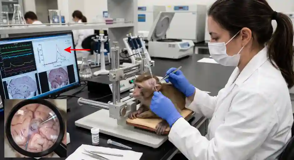
- Optics provides the control. A thin optical fiber delivers pulses of light to the brain region of interest, and the targeted cells respond.
Deisseroth’s group and others soon added more tools. They developed channels that switch neurons off with light and variants that stay active after a brief flash. They also built methods to deliver light and genes in living, freely moving animals.
What it has taught us
Optogenetics has revealed how specific neurons and neural pathways govern behavior, such as eating, sociability and aggression.
- Sleep and waking: activating a small group of neurons that make the arousal chemical hypocretin wakes sleeping mice.
- Parkinson’s disease: stimulating particular circuits in the basal ganglia relieved movement symptoms in animal models, which helped researchers understand why deep brain stimulation works.
Researchers who use it to study memory, fear, addiction, depression and many other conditions can now ask a causal question. Instead of merely observing that a cell is active during a behavior, they can turn that cell on or off and see whether the behavior changes.
From the lab to the clinic: restoring vision
The Nobel committee noted that researchers have also taken the first steps toward using optogenetics as a medical treatment, and there are hopes that it could improve cochlear implants. The most visible clinical effort is in blindness.
What goes wrong in retinitis pigmentosa (RP). In RP, an inherited disease, the photoreceptors in the retina (the rods and cones that catch light) gradually die. People first lose night and side vision and may eventually lose almost all sight. Mutations in more than 100 different genes can cause RP, which makes gene-by-gene treatments difficult.
The optogenetic idea. Although the photoreceptors are gone, other retinal cells that normally pass signals to the brain often survive. As Per Svenningsson of the Karolinska Institute explained, researchers use optogenetics to stimulate these remaining healthy cells, activating the optic nerve and generating visual perception in the brain. A gene therapy delivers the gene for a light-sensitive protein to those surviving cells. They become new, makeshift light detectors. This does not “override” the retina so much as substitute for the missing photoreceptors, and the approach does not depend on which gene caused the disease. Early animal work showed the principle. In 2021, researchers reported the first partial recovery of visual function in a blind patient, a person with RP who used special light-amplifying goggles alongside the therapy.
MOGENRY (MCO-010). Nanoscope Therapeutics’ therapy, sonpiretigene isteparvovec, carries a gene for a “multi-characteristic opsin.” It is delivered by an engineered virus (AAV) in an injection into the eye’s gel-like interior. The company said the therapy is designed to respond to ambient light. The FDA has accepted its biologics license application for adults with RP and severe vision loss, and the filing is supported by data from the RESTORE trial. One industry tracker lists the review’s target action date as June 30, 2027. If approved, it would be positioned as the first mutation-agnostic therapy intended to restore useful vision in RP regardless of the causative gene.
A caution is in order. MOGENRY is still under review and not approved. The vision it restores is partial, closer to detecting shapes, light and motion than to normal sight, and long-term safety and durability are still being studied. Readers should not treat any optogenetic therapy as a cure yet.
Why this prize matters
This Nobel is an unusual case of a discovery that began with curiosity about pond algae and ended up reshaping neuroscience. Hegemann and Nagel found the molecular switch, and Deisseroth showed how to use it in the brain. As Nobel Assembly Secretary-General Thomas Perlmann put it, “We can get a whole new understanding of the function of the brain.” Others contributed to the field too, including Ed Boyden and Feng Zhang, who worked with Deisseroth on the early neuron experiments, and Gero Miesenböck, who pioneered an earlier light-based approach. A Nobel Prize can honor at most three people.
References
Adamantidis, A. R., Zhang, F., Aravanis, A. M., Deisseroth, K., & de Lecea, L. (2007). Neural substrates of awakening probed with optogenetic control of hypocretin neurons. Nature, 450(7168), 420–424. https://doi.org/10.1038/nature06310
Berndt, A., Yizhar, O., Gunaydin, L. A., Hegemann, P., & Deisseroth, K. (2009). Bi-stable neural state switches. Nature Neuroscience, 12(2), 229–234. https://doi.org/10.1038/nn.2247
Bi, A., Cui, J., Ma, Y.-P., Olshevskaya, E., Pu, M., Dizhoor, A. M., & Pan, Z.-H. (2006). Ectopic expression of a microbial-type rhodopsin restores visual responses in mice with photoreceptor degeneration. Neuron, 50(1), 23–33. https://doi.org/10.1016/j.neuron.2006.02.026
Boyden, E. S., Zhang, F., Bamberg, E., Nagel, G., & Deisseroth, K. (2005). Millisecond-timescale, genetically targeted optical control of neural activity. Nature Neuroscience, 8(9), 1263–1268. https://doi.org/10.1038/nn1525
Chow, B. Y., Han, X., Dobry, A. S., Qian, X., Chuong, A. S., Li, M., Henninger, M. A., Belfort, G. M., Lin, Y., Monahan, P. E., & Boyden, E. S. (2010). High-performance genetically targetable optical neural silencing by light-driven proton pumps. Nature, 463(7277), 98–102. https://doi.org/10.1038/nature08652
Crick, F. H. C. (1979). Thinking about the brain. Scientific American, 241(3), 219–232.
Deisseroth, K. (2015). Optogenetics: 10 years of microbial opsins in neuroscience. Nature Neuroscience, 18(9), 1213–1225. https://doi.org/10.1038/nn.4091
Foster, K. W., Saranak, J., Patel, N., Zarrilli, G., Okabe, M., Kline, T., & Nakanishi, K. (1984). A rhodopsin is the functional photoreceptor for phototaxis in the unicellular eukaryote Chlamydomonas. Nature, 311(5988), 756–759. https://doi.org/10.1038/311756a0
Gradinaru, V., Mogri, M., Thompson, K. R., Henderson, J. M., & Deisseroth, K. (2009). Optical deconstruction of parkinsonian neural circuitry. Science, 324(5925), 354–359. https://doi.org/10.1126/science.1167093